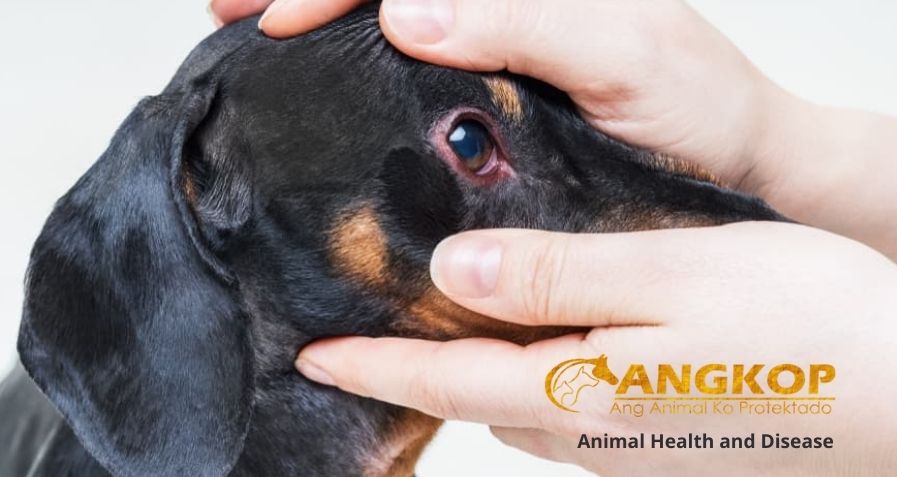
Red Eye
Issues
Red Eye
Red Eye – hyperemia of the eyelids or ocular vasculature, or hemorrhage within the eye.
Active dilation of ocular vessels—in response to extraocular or intraocular inflammation or passive congestion.
Hemorrhage from existing or newly formed blood vessels.
SYSTEMS AFFECTED
Ophthalmic—eye and/or ocular adnexa
SIGNALMENT
Dog and cat
SIGNS
Historical Findings
- Depends on cause
Physical Examination Findings
- Depends on cause.
- May affect one or both eyes.
- Result of systemic disease—abnormalities in other organ systems common.
CAUSES
- Virtually every case fits into one or more of the following categories.
- Blepharitis.
- Conjunctivitis.
- Keratitis.
- Episcleritis or scleritis.
- Anterior uveitis.
- Glaucoma.
- Hyphema.
- Orbital disease—usually the orbital abnormality is more prominent.
RISK FACTORS
- Systemic infectious or inflammatory diseases.
- Immunocompromise.
- Coagulopathies.
- Systemic hypertension.
- Irritation by potentially any topical ophthalmic medication.
- Neoplasia.
- Trauma.
DIAGNOSIS
DIFFERENTIAL DIAGNOSIS
More than one cause may occur simultaneously.
Similar Signs
- Rule out normal variations.
- Palpebral conjunctiva—normally redder than bulbar conjunctiva.
- One or two large episcleral vessels—may be normal if the eye is otherwise quiet.
- Transient mild hyperemia—with excitement, exercise, and straining.
- Horner’s syndrome—may cause mild conjunctival vascular dilation; differentiated by other signs and pharmacologic testing.
Causes
- Superficial (conjunctival) vessels—originate near the fornix; move with the conjunctiva; branch repeatedly; blanch quickly with topical 2.5% phenylephrine or 1:100,000 epinephrine; suggest ocular surface disorders (e.g., conjunctivitis, superficial keratitis, blepharitis).
- Deep (episcleral) vessels—originate near the limbus; branch infrequently; do not move with the conjunctiva; blanch slowly or incompletely with topical sympathomimetics; suggest episcleritis or intraocular disease (e.g., anterior uveitis or glaucoma).
- Discharge—mucopurulent to purulent: typical of ocular surface disorders and blepharitis; serous or none: typical of intraocular disorders.
- Swollen or inflamed eyelids—suggest blepharitis.
- Corneal opacification, neovascularization, or fluorescein stain retention—suggest keratitis.
- Aqueous flare or cell (increased protein or cells in the anterior chamber)—confirms diagnosis of anterior uveitis.
- Pupil—miotic: common with anterior uveitis; dilated: common with glaucoma; normal: with blepharitis and conjunctivitis.
- Abnormally shaped or colored irides—suggest anterior uveitis.
- Luxated or cataractous lenses—suggest glaucoma or anterior uveitis.
- IOP—high: diagnostic for glaucoma; low: suggests anterior uveitis.
- Loss of vision—suggests glaucoma, anterior uveitis, or severe keratitis.
- Glaucoma and anterior uveitis—may complicate hyphema.
CBC/BIOCHEMISTRY/URINALYSIS
Typically, normal, except with anterior uveitis, glaucoma, or hyphema secondary to systemic disease.
See Anterior Uveitis—Dogs; Anterior Uveitis—Cats; Hyphema.
OTHER LABORATORY TESTS
Depends on cause
IMAGING
- Chest radiographs—consider with anterior uveitis or if intraocular neoplasia is a possibility.
- Abdominal radiography or ultrasonography—may help rule out infectious or neoplastic causes.
- Ocular ultrasonography—if the ocular media are opaque; may define the extent and nature of intraocular disease or identify an intraocular tumor.
DIAGNOSTIC PROCEDURES
- Tonometry—must perform in every patient with an unexplained red eye.
- Ocular Surface Disorders
- Aerobic bacterial culture and sensitivity profile—with a purulent discharge, chronic disease, or if the response to treatment is poor.
- Schirmer tear test.
- Cytologic examination of affected tissue—lid; conjunctiva; cornea.
- Cats—consider PCR or IFA test on corneal or conjunctival scrapings for feline herpesvirus and Chlamydia; collect sample before fluorescein staining to avoid false-positive results on IFA.
- Fluorescein stain.
- Conjunctival biopsies—with chronic conjunctivitis or with a mass lesion.
- See specific disease—Conjunctivitis—Cats, Conjunctivitis—Dogs; Blepharitis; Keratitis chapters.
- Intraocular Disorders
- Fluorescein stain.
- See specific disease—Anterior Uveitis—Cats; Anterior Uveitis—Dogs; Hyphema; Glaucoma.
TREATMENT
- Usually outpatient.
- Elizabethan collar—considered to prevent self-trauma.
- Avoid dirty environments or those that may lead to ocular trauma, especially if topical corticosteroids are used.
- Because there is a narrow margin for error, consider referral if you cannot attribute the condition to one of the listed causes, if you cannot rule out glaucoma on the initial visit, or if the diagnosis is so uncertain that administration of a topical antibiotic alone or a topical corticosteroid alone would be questionable.
- Few causes are fatal; however, a workup may be indicated (especially with anterior uveitis and hyphema) to rule out potentially fatal systemic diseases.
- Deep corneal ulcers and glaucoma—may be best treated surgically.
MEDICATIONS
DRUG(S)
- Depends on specific cause.
- Generally, control ocular pain, inflammation, infection, and IOP.
- Aspirin 10–15 mg/kg PO q8–12h (dogs); may control mild ocular inflammation and pain pending test results.
- Carprofen 2.2 mg/kg q12h or 4.4 mg/kg q24h.
- Flunixin meglumine 0.5 mg/kg IV once; may be used in dogs with severe ocular inflammation pending test results.
- CONTRAINDICATIONS
- Topical corticosteroids—contraindicated if the cornea retains fluorescein stain.
- Systemic corticosteroids—avoid until infectious systemic causes have been ruled out.
PRECAUTIONS
Topical aminoglycosides—may be irritating; may impede reepithelization if used frequently or at high concentrations.
Topical solutions—may be preferable to ointments if corneal perforation is possible.
Atropine—may exacerbate KCS and glaucoma.
NSAIDs—use with caution in hyphema.
FOLLOW-UP
PATIENT MONITORING
- Depends on cause.
- Repeat ophthalmic examinations—as required to ensure that IOP, ocular pain, and inflammation are well controlled.
- The greater the risk of loss of vision, the more closely the patient needs to be followed; may require daily or more frequent examination.
POSSIBLE COMPLICATIONS
- Death
- Loss of the eye or permanent vision loss
- Chronic ocular inflammation and pain
MISCELLANEOUS
ASSOCIATED CONDITIONS
Numerous systemic diseases
ZOONOTIC POTENTIAL
See Anterior Uveitis—Dogs; Anterior Uveitis—Cats; Conjunctivitis—Dogs; Conjunctivitis—Cats; Episcleritis; Glaucoma; Keratitis, Ulcerative
PREGNANCY/FERTILITY/BREEDING
Systemic corticosteroids may complicate pregnancy.
ABBREVIATIONS
IFA = immunofluorescent antibody
IOP = intraocular pressure
KCS = keratoconjunctivitis sicca
NSAID = nonsteroidal anti-inflammatory drug
PCR = polymerase chain reaction
Visit your veterinarian as early recognition, diagnosis, and treatment are essential.
You may also visit https://www.facebook.com/angkopparasahayop