Roundworms (Ascariasis)
Issues
Roundworms (Ascariasis)
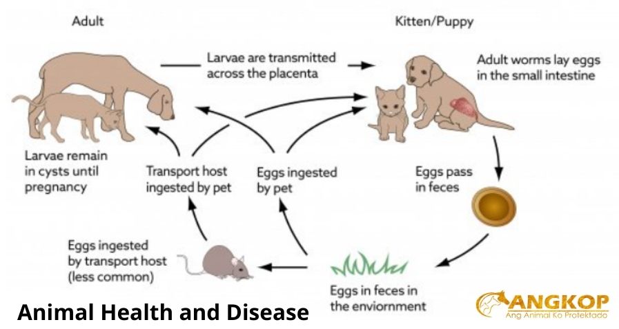
Ascariasis caused by Toxocara canis (dogs), T. cati (cats), and Toxascaris leonina (dogs and cats); Baylisascaris (raccoons) can infect dogs and cause neurocysticercosis in people.
- Transplacental transmission of T. canis larvae from bitch’s tissues to pups causes prenatal infection; transmammary transmission of larvae occurs with both Toxocara spp.; no transplacental or transmammary transmission occurs with Toxascaris.
- In the first month of life, infected neonatal pups may develop abdominal pain and rapidly deteriorate before eggs appear in feces.
- Older pups and kittens can acquire infection by ingesting eggs disseminated on premises by dams with post-gestational infection; dams can be infected by ingesting immature stages in pups’ feces or vomitus, or by predation on vertebrate transport hosts.
- Adult ascarids occur in the lumen of the small intestine; larval stages of Toxocara spp. may migrate in the liver and lungs.
- If very numerous, ascarids (up to 10–12 cm long) can distend intestine leading to colic, interference with gut motility, inability to utilize food, and intestinal rupture.
SIGNALMENT
Dog and cat.
- Important clinically in pups and kittens due to transmission in utero and/or in colostrum/milk.
SIGNS
- Abdominal distension (“pot belly”); often with palpable intestinal distension.
- Colic.
- Weakness, loss of condition, cachexia; poor nursing or appetite; scant feces.
- Coughing due to larval lung migration.
- Entire litter may be affected.
CAUSES & RISK FACTORS
- Toxocara infection of bitch with dormant larvae in tissues or infection of queen during late pregnancy or early lactation.
- Access to infected transport hosts.
- Concurrent enteric infections.
- Larvae undergo extensive migrations throughout the body and can cause granulomatous inflammation (visceral larva migrans). Visceral larva migrans caused by a variety of ascarids is a major cause of morbidity in humans throughout the world.
DIAGNOSIS
DIFFERENTIAL DIAGNOSIS
- Transmammary perinatal infection of neonates with hookworms (anemia, melena, weakness, lethargy, pale mucous membranes, enteritis) or Strongyloides (diarrhea); coccidiosis, giardiasis also cause enteritis in pups and kittens; examine feces to identify eggs or larvae.
- Physaloptera can also occur in vomitus.
CBC/BIOCHEMISTRY/URINALYSIS
Usually normal
DIAGNOSTIC PROCEDURES
Fecal flotation to detect eggs; Toxocara eggs spherical pitted outer shell membrane, single dark cell filling interior, 80–85 μm (T. canis), ∼75 μm (T. cati).
Baylisascaris eggs are similar to T. canis eggs but smaller (∼76 × 60 μm), more finely pitched shell.
Toxascaris eggs are ovoid, smooth exterior shell membrane, 1–2 cells with light-colored cytoplasm; cells do not fill the interior of the egg; 80 × 70 μm in diameter.
Necropsy of siblings that died of similar signs.
Identify ascaris in feces, vomitus, or small intestine by size, presence of three lips, and cervical alae.
TREATMENT
Anthelmintic treatment as outpatient.
Acute severe cases treated as inpatients; supplement with intravenous fluids.
Milbemycin or other anthelmintic effective against T. canis suggested for treatment (extra-label) of Baylisascaris in dogs.
Alert client to possibility of sudden death or chronic debilitation.
Treat bitch or queen with adulticide/larvicide anthelmintic to remove intestinal stages and to limit transmission to subsequent litters.
PATIENT MONITORING
Repeat post-treatment fecal exams on pups/kittens and/or repeat anthelmintic treatment every 2–3 weeks until old enough for monthly anthelmintic product because migrating larvae acquired from bitch or dam will continue to mature.
PREVENTION/AVOIDANCE
To minimize environmental contamination with infective eggs, treat infected dogs/cats with anthelmintic and promptly dispose of feces.
Prevent dogs/cats from hunting or ingesting transport hosts.
Extra-label treatment of bitch or queen with adulticide/larvicide anthelmintic to remove intestinal stages and decrease vertical transmission to offspring.
POSSIBLE COMPLICATIONS
Transplacental transmission of large numbers of larvae can result in fetal death or birth of weak, non-viable pups.
Infection with large numbers of ascarids can cause blockage, possible rupture of small intestine.
EXPECTED COURSE AND PROGNOSIS
Prognosis good after anthelmintic treatment; guarded with severe prenatal T. canis infection.
MISCELLANEOUS
AGE-RELATED FACTORS
Greater clinical concern in neonates
ZOONOTIC POTENTIAL
Visceral larva migrans, ocular larva migrans, neural larva migrans, chronic abdominal or cutaneous problems may follow ingestion of infective Toxocara spp. or Baylisascaris eggs by humans.
Most likely cause of neural larva migrans is Baylisascaris. Virtually all raccoons become infected with Baylisascaris and therefore extreme caution should be exercised with clients having raccoons as “pets.”
Visit your veterinarian as early recognition, diagnosis, and treatment are essential.
You may also visit – https://www.facebook.com/angkopparasahayop