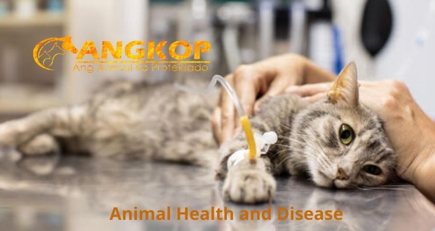
Pancreatitis—Cats

Issues
Pancreatitis—Cats
Cats can suffer from two forms of pancreatitis: acute and chronic. Clinical signs can be similar for either form but tend to come on more rapidly and are more severe in cats with acute pancreatitis. The most common clinical signs are very vague, including lethargy and a reduced appetite. About 50% of cats will have vomiting or weight loss, and some cats will develop diarrhea as well. Abdominal pain, while a very common finding in human and canine pancreatitis, is only reported in about 10-30% of cats with pancreatitis, but this may be related to cats’ stoic nature and ability to hide signs of pain from their owners and veterinarians. Some cats with chronic pancreatitis may show very mild or almost unnoticeable signs, while cats with severe, acute pancreatitis can become suddenly critically ill.
Inflammation of the pancreas is most often of unknown cause(s).
- Acute pancreatitis – inflammation of the pancreas that occurs abruptly with little or no permanent pathologic change.
- Chronic pancreatitis – continuing inflammatory disease that is accompanied by irreversible morphologic change such as fibrosis.
PATHOPHYSIOLOGY
- Host defense mechanisms normally prevent pancreatic autodigestion by pancreatic enzymes, but under select circumstances, these natural defenses fail; autodigestion occurs when these digestive enzymes are activated within acinar cells.
- Local and systemic tissue injury is due to the activity of released pancreatic enzymes and a variety of inflammatory mediators such as kinins, free radicals, and complement factors are released by infiltrating neutrophils and macrophages. The most common pathologies involving the feline pancreas include acute necrotizing pancreatitis (ANP) and acute suppurative pancreatitis.
SYSTEMS AFFECTED
- Gastrointestinal – altered GI motility (ileus) due to regional chemical peritonitis; local or generalized peritonitis due to enhanced vascular permeability; concurrent inflammatory bowel disease may be seen in some cats.
- Hepatobiliary – lesions due to shock, pancreatic enzyme injury, inflammatory cellular infiltrates, hepatic lipidosis, and intra/extrahepatic cholestasis. Feline gastrointestinal inflammatory disease (concurrent cholangitis ± inflammatory bowel disease) may be seen in some cats.
- Respiratory – pulmonary edema or pleural effusion.
- Cardiovascular—cardiac arrhythmias may result from release of myocardial depressant factor.
- Hematologic – activation of the coagulation cascade and systemic consumptive coagulopathy (DIC) occur.
GENETICS
No genetic basis for disease pathogenesis in cats has been identified.
INCIDENCE/PREVALENCE
True prevalence is unknown but is a relatively common clinical disorder in cats.
Necropsy surveys suggest an increased prevalence in cats with cholangitis, and inflammatory bowel disease. The unique feline pancreaticobiliary anatomy and intestinal microbiota likely contribute to multi-organ inflammatory disease in this species.
GEOGRAPHIC DISTRIBUTION
Worldwide
SIGNALMENT
Species
Cat of any age
Breed Predilections
Siamese cats
Mean Age and Range
Mean age for acute pancreatitis is 7.3 years.
Predominant Sex
None
SIGNS
General Comments
Vague, nonspecific, and nonlocalizing signs. Anorexia, lethargy, and vomiting are reported most frequently.
Historical Findings
- Lethargy/anorexia
- Vomiting
- Weakness
- Abdominal pain
- Diarrhea – small bowel and large bowel diarrhea and fever are less common in cats than dogs
- Physical Examination Findings
- Severe lethargy.
- Dehydration—common; due to GI losses.
- Abdominal pain—may adopt a “prayer position” and/or resist abdominal palpation. Abdominal pain is recognized much less frequently in cats compared to dogs.
- Mass lesions may be palpable.
- Fever—Observed in 25% of cats.
CAUSES
- Etiology is most often unknown; possibilities include:
- Hepatobiliary tract disease—both inflammatory and degenerative (hepatic lipidosis)
- Pancreatic trauma/ischemia
- Duodenal reflux
- Drugs/toxins (organophosphates)
- Pancreatic duct obstruction
- Hypercalcemia
- Inflammatory gastrointestinal disease
- Nutrition—excessive lean body mass is associated with ANP
DIAGNOSIS
DIFFERENTIAL DIAGNOSIS
- Other causes of acute abdomen.
- GI disease (obstruction, foreign body, perforation, infectious gastroenteritis, ulcer disease)—exclude with CBC/biochemistry/urinalysis, diagnostic imaging, and paracentesis. Gastrointestinal or hepatic neoplasia—exclude with tissue biopsy.
- Urogenital disease (pyelonephritis, prostatitis or abscessation, pyometra, urinary tract rupture or obstruction, acute renal failure)—exclude with CBC/biochemistry/urinalysis, urine culture/sensitivity, and imaging.
- Hepatobiliary disease (cholangitis and EHBO) excludes with CBC/biochemistry/urinalysis, bile acids, imaging, and liver biopsy.
- Abdominal neoplasia—exclude with imaging and cytology or biopsy.
CBC/BIOCHEMISTRY/URINALYSIS
- CBC—often reveals nonregenerative anemia (40%), leukocytosis (38%), and/or leukopenia (15%).
- Serum biochemistries—often show prerenal azotemia; liver enzyme activities (ALT, ALP) are often elevated because of hepatic ischemia or exposure to pancreatic enzymes; hyperbilirubinemia with intra/extrahepatic biliary obstruction; hyperglycemia with necrotizing pancreatitis due to hyperglucagonemia; hypoalbuminemia, hypercholesterolemia and hypertriglyceridemia are common. Hypocalcemia is more common in cats than dogs, and a low ionized calcium concentration is a negative prognostic indicator in cats.
- Urinalysis—increased urine SG associated with dehydration or can be unremarkable.
OTHER LABORATORY TESTS
- Serum amylase and lipase activities are unreliable serologic markers—may be elevated, but are nonspecific; can also increase with hepatic, renal, or neoplastic disease in the absence of pancreatitis.
- Serum pancreatic lipase immunoreactivity (fPL) is a highly sensitive and specific serologic marker of acute pancreatic inflammation. A cage-side fPL assay (SNAP fPL) has been developed as a useful screening tool. Elevation in SNAP fPL should be followed up by laboratory measurement of serum Spec fPL to quantitate the degree of elevation.
- IMAGING
- Abdominal radiographs—may include increased soft tissue opacity in the right cranial abdominal compartment; loss of visceral detail (“ground glass appearance”) due to abdominal effusion; static gas pattern in the proximal duodenum.
- Abdominal ultrasound—nonhomogeneous solid or cystic mass lesions suggest pancreatic abscess; may be a pancreatic mass or altered echogenicity (hypoechoic) in the area of the pancreas; pancreas is usually enlarged with irregular borders, surrounding mesentery may be hyperechoic due to focal peritonitis, may see peritoneal effusion and extrahepatic biliary obstruction.
- fPL assay and pancreatic ultrasound in combination have the highest specificity for an antemortem diagnosis of acute pancreatitis.
DIAGNOSTIC PROCEDURES
- Ultrasound-guided needle-aspiration biopsy may confirm inflammation (cytology), abscess, or cyst.
- Laparoscopy with pancreatic forceps biopsy for histologic diagnosis.
- Histopathologic evaluation may miss focal or segmental pancreatic inflammation and results should be interpreted with caution.
PATHOLOGIC FINDINGS
- Gross findings (acute pancreatitis)—mild swelling with edematous pancreatitis.
- Gross findings (chronic pancreatitis)—pancreas is reduced in size, firm, gray, and irregular; may contain extensive adhesions to surrounding viscera.
- Microscopic changes (acute pancreatitis)—include edema, parenchymal necrosis, hemorrhage, and neutrophilic cellular infiltrate with acute lesions.
- Microscopic changes (chronic pancreatitis)—pancreatic fibrosis around ducts, ductal epithelial hyperplasia, atrophy, and mononuclear cellular infiltrate.
TREATMENT
APPROPRIATE HEALTH CARE
- Inpatient medical management most often.
- Aggressive IV fluid therapy.
- Fluid therapy goals—correct hypovolemia and maintain pancreatic microcirculation.
- A balanced electrolyte solution such as lactated Ringer’s solution (LRS) is the first-choice rehydration fluid.
- Correct initial dehydration (mL = % dehydration × weight in kg × 1,000) and give over 4–6 hours.
- May need colloids (oxyglobin, hetastarch) to improve pancreatic circulatory needs and prevent ischemia.
- Following replacement of deficits, give additional fluids to match maintenance requirements (2.5 × weight in kg) and ongoing losses (estimated).
- Potassium chloride (KCl) supplementation usually needed because of potassium loss in the vomitus; base potassium supplementation on measured serum levels (use 20 mEq of KCl/L of IV fluid if serum potassium levels are not known; do not administer faster than 0.5 mEq/kg/h).
ACTIVITY
Restrict
DIET
- Continue to feed orally unless vomiting is intractable; feeding maintains intestinal epithelial integrity and minimizes bacterial translocation.
- Initiate enteral feeding via esophagostomy, gastrostomy enteral feeding device, or nasoesophageal tube placement.
- NPO in animals with persistent vomiting for the shortest time possible; when there has been no vomiting for 12 hours, offer small volumes of water; if tolerated, begin small, frequent feedings of a diet that does not contain excessive amounts of dietary fat. Most nutritionists agree that excessive dietary fat restriction is not necessary in cats with pancreatitis.
CLIENT EDUCATION
- Discuss the need for extended hospitalization.
- Discuss the expense of diagnosis and treatment.
- Discuss possible short-term and long-term complications.
SURGICAL CONSIDERATIONS
- May need surgery to remove pseudocysts, abscesses, or devitalized tissue seen with necrotizing pancreatitis.
- May need laparotomy and pancreatic biopsy to confirm pancreatitis and/or rule out other, nonpancreatic diseases such as hepatic cholangitis, lipidosis, and/or IBD.
- Extrahepatic biliary obstruction from pancreatitis requires ductal decompression with surgical correction.
PREVENTION/AVOIDANCE
- Weight reduction if obese
- Avoid high-fat diets
POSSIBLE COMPLICATIONS
- Failed response to supportive therapy.
- Life-threatening associated conditions such as EPI, diabetes mellitus, and hepatic lipidosis.
- Progression of acute pancreatitis to chronic pancreatitis.
EXPECTED COURSE AND PROGNOSIS
- Guarded for most patients with ANP; cats with multi-organ inflammation may be less responsive to treatment.
- More guarded to poor for patients with severe necrotizing pancreatitis, decreased ionized calcium fraction, and systemic conditions.
MISCELLANEOUS
ASSOCIATED CONDITIONS
Life-Threatening
- Pulmonary edema (e.g., adult respiratory distress syndrome)
- Cardiac arrhythmias
- Peritonitis
- DIC
Non-Life-Threatening
- Diabetes mellitus
- EPI
- Chronic pancreatitis
- Cholangitis and hepatic lipidosis
- Inflammatory bowel disease
ABBREVIATIONS
ALP = alkaline phosphatase
ALT = alanine aminotransferase
ANP = acute necrotizing pancreatitis
DIC = disseminated intravascular coagulation
EHBO = extrahepatic biliary obstruction
EPI = exocrine pancreatic insufficiency
fPL = feline pancreatic lipase immunoreactivity
GI = gastrointestinal
IBD = inflammatory bowel disease
NPO = nothing per os
Visit your veterinarian as early recognition, diagnosis, and treatment are essential.
You may also visit – https://www.facebook.com/angkopparasahayop
