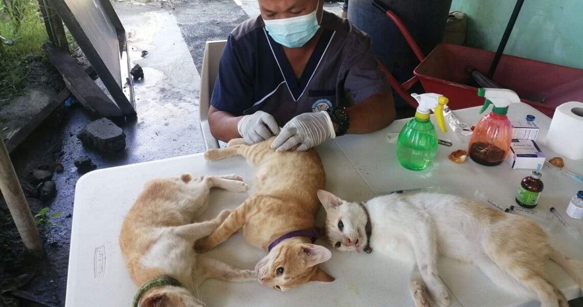
Brain Injury
There are many ways dogs and cats can sustain brain injuries. Common causes of brain injury in pets include being hit by a vehicle, attacks by larger animals (e.g. being severely shaken and/or bitten), falling from a high height, blunt force trauma, or gunshot wounds.
Brain injuries can be classified as traumatic, caused by external forces or Non-traumatic, caused by non-violent forces (e.g., hypoxia, metabolic disorders, vascular disruption, infection, toxicity, neoplasia).
It can be primary, a direct initial insult when tissue and vessels are stretched, compressed or torn or secondary, alterations of brain vasculature and tissue following primary injury.
PATHOPHYSIOLOGY
Acceleration, deceleration, and rotational forces traumatize brain tissue.
High oxygen and glucose requirements put the brain at risk for hypoxia.
Oxygen delivery is dependent on CBF and CPP (= MAP – ICP).
Intracranial bleeding, edema (vasogenic and cytotoxic), vasodilation, and/or vasospasms increase ICP, causing low CBF, ischemia, brain swelling, and herniation; slow, progressive increase in ICP better tolerated than small, acute rise.
Hypotension, hypoxia—major contributors to secondary injury.
SYSTEMS AFFECTED
Nervous—altered mentation, cranial nerve deficits, seizures, twitching, postural changes.
Cardiovascular—arrhythmias.
Endocrine/Metabolic—alterations in ADH release and sodium concentration; central temperature dysregulation; insulin resistance; depletion of cortisol.
Ophthalmic—changes in eye position, eye movements, pupillary light reflexes, papilledema.
Respiratory—hyper- and hypocapnea; abnormal breathing patterns; neurogenic pulmonary edema.
INCIDENCE/PREVALENCE
Head and neck injuries are found in up to 34% of dogs and cats suffering blunt force trauma.
Parenchymal and extradural hematomas are found in 10% of dogs and cats with signs of mild head injury and in up to 80% with a severe head injury.
SIGNS
Historical Findings
Determine cause—trauma; cardiac arrest; heart failure; hypertension; toxins; coagulopathies; severe respiratory compromise; prolonged seizures; hypoglycemia.
The decline in neurologic condition—implies a progression from intracranial bleeding, cerebral edema, ischemia.
Seizure activity—cerebral or diencephalon involvement.
Physical Examination Findings
Evidence of head trauma—open wounds, epistaxis, blood in the ear canals.
Cardiac or respiratory insufficiency—hypoxia, cyanosis, hypoventilation.
Poor perfusion—weak pulse, pale mucous membranes.
Skull palpation—fractures, open fontanelles.
Sustained bradycardia—midbrain, pontine, or medullary lesion.
Cushing’s reflex—bradycardia and hypertension.
Ecchymosis, petechiae, retinal hemorrhages or distended vessels—hypertension, coagulopathy.
Papilledema—cerebral edema.
Retinal detachment—infectious, neoplastic, or hypertensive causes.
Neurologic Examination Findings
Mental Status
Level of consciousness and cranial nerve deficits—localize the lesion to the cerebral cortex (better prognosis), midbrain/brainstem, or multifocal.
Postural changes—decerebrate rigidity with midbrain lesion; decerebellate rigidity with the cerebellar lesion.
Peracute focal deficits suggest vascular or neoplastic causes.
Pupillary Light Reflexes
Miotic responsive pupils—cerebral or diencephalic lesion (rule out traumatic uveitis, Horner’s syndrome).
Pinpointed unresponsive pupils—diencephalic, pontine, or medullary lesion.
Dilated unresponsive pupil(s) or midpoint fixed unresponsive pupils—midbrain lesion.
Cranial Nerves
Normal with altered mentation—cerebrum-diencephalon lesion.
CN II- Loss of menace and dazzle response with dilated unresponsive pupils—cranial forebrain.
Loss of physiologic nystagmus—brainstem lesion
CN III—midbrain lesion.
CN V–XII—pontine or medullary lesion.
Respiratory Patterns
Cheyne-Stokes—severe diffuse cerebral or diencephalon lesion
Hyperventilation—midbrain lesion
Ataxic or apneustic—pontine or medullary lesion
CAUSES
Trauma
Prolonged hypoxia or ischemia
Prolonged shock
Severe hypoglycemia
Prolonged seizures
Severe hyperthermia or hypothermia
Alterations in serum osmolality
Toxins
Neoplasia
Hypertension
Hemorrhage
Inflammatory, infectious, immune-mediated diseases
Thiamin deficiency
Hydrocephalus
Parasitic migration
RISK FACTORS
Free-roaming—trauma, toxins
Coexisting cardiac, respiratory, hemostatic, hepatic disease
Diabetes mellitus—insulin therapy
DIFFERENTIAL DIAGNOSIS
Systemic causes of altered states of consciousness or central vestibular signs—metabolic disease; toxins; drugs; infection.
CBC/BIOCHEMISTRY/URINALYSIS
Reflect systemic effects of neurologic signs
Alterations in serum sodium suggest central ADH abnormalities
OTHER LABORATORY TESTS
Arterial blood gas
Coagulation profile.
Infectious disease titers
IMAGING
Skull radiographs—detect fractures.
CT—detects acute hemorrhage, infarcts, fractures, penetrating foreign bodies, hydrocephalus, herniation.
MRI—detects cerebral edema, hemorrhage, mass, hydrocephalus, infiltrative diseases, inflammation, herniation, fractures.
Ultrasound optic disk—if > 3 mm diameter, may be associated with brain edema.
DIAGNOSTIC PROCEDURES
ECG—detects arrhythmias
BP—determine perfusion
CSF analysis—if cause unknown and no contraindications
PATHOLOGIC FINDINGS
Brain edema or inflammation
Herniation
Hemorrhage
Hydrocephalus
Infarct
Laceration, contusion
Hematomas
Skull fracture
Necrosis
Apoptosis
TREATMENT
APPROPRIATE HEALTH CARE
Goals of therapy—maximize oxygenation and ventilation; support BP and CPP; decrease ICP; decrease cerebral metabolic rate.
Maintain systolic BP > 90 mmHg and PCO2 at 35–40 mm Hg; with suspected elevated ICP, hyperventilation to 32–35 mmHg.
Maintain PaO2 > 60 mmHg, SaO2 > 90%, SpO2 > 94%.
Avoid cough or sneeze reflex during intubation or nasal oxygen supplementation; lidocaine (dogs: 1–2 mg/kg IV) before.
Do not compress jugular veins.
Orotracheal intubation if gag reflex lost.
NURSING CARE
Aggressive therapy for midbrain/brainstem lesion or declining neurologic signs.
Overzealous fluid resuscitation can contribute to brain edema.
Small-volume fluid resuscitation techniques to maintain systolic BP > 90 mmHg with normal heart rate.
Combination of isotonic crystalloids (10–20 mL/kg increments) with hydroxyethyl starch (5 mL/kg increments) over 5–8 minutes.
Avoid hypertension.
Level head with body or elevate head and neck to a 20° angle.
Keep airway unobstructed; use suction and humidify if intubated; hyper oxygenate and consider IV lidocaine prior to suctioning.
Lubricate eye.
Reposition every 2–4 hours to avoid hypostatic pulmonary congestion.
Prevent fecal/urine soiling.
Maintain normal core body temperature.
Maintain hydration with a balanced electrolyte crystalloid solution.
Rehabilitation exercises.
ACTIVITY
Restricted.
Consult rehabilitation specialists for appropriate exercises to maintain muscle tone.
DIET
Initiate trickle flow feeding to meet elevated metabolic demands.
CLIENT EDUCATION
Neurologic signs may worsen before improving.
Neurologic recovery may not be evident for several days; possibly > 6 months for residual neurologic deficits.
Serious systemic abnormalities contribute to CNS instability.
SURGICAL CONSIDERATIONS
Depressed skull fracture, penetrating the foreign body, uncontrollable ICP elevation (insufficient CSF drainage, hematoma/mass evacuation, herniation).
MEDICATIONS
DRUG(S) OF CHOICE
Elevated ICP
Ensure systolic BP > 90 mmHg. Lower ICP by hyperventilation, drug therapy, drainage of CSF from the ventricles, or surgical decompression.
7% hypertonic saline—2–4 mL/kg IV; can reduce fluid volume needed to reach resuscitation endpoints; combine with colloid.
Furosemide—0.75 mg/kg IV; may decrease CSF production; used in patients with congestive heart failure, volume overload, hyperosmolar diseases, or anuric renal failure; use before mannitol.
Mannitol—0.1–0.5 g/kg IV bolus repeated at 2-hour intervals 3–4 times in dogs, and 2–3 three times in cats; repeated doses must be given on time; improves brain blood flow and lowers ICP; may exacerbate hemorrhage.
Glucocorticosteroids—no benefit in acute management and long-term outcome in humans; higher morbidity. Anti-inflammatory doses (prednisone 1 mg/kg/day) may be of benefit with brain edema related to intracranial neoplasia and infectious meningitis. Immunosuppressive doses (2 mg/kg/day) in combination with additional immunosuppressive drugs in immune-mediated meningitis.
Provide analgesia/sedatives (e.g., fentanyl 3–5 μg/kg IV then 3–5 μg/kg/h CRI ± lidocaine 3–5 mg/kg/h) as indicated. Avoid agents that can reduce CPP.
Thrashing, seizures, or uncontrolled motor activity—diazepam CRI (0.5–1 mg/kg/h), midazolam CRI (0.2–0.4 mg/kg IV), or propofol (3–6 mg/kg IV titrated to effect; 0.1–0.6 mg/kg/min CRI) monitor for hypotension; intubate if unable to protect airway.
Levetiracetam 20 mg/kg IV/IM/rectal q8h if seizure activity.
Other
Reducing cerebral metabolic rate with heavy sedation using dexmedetomidine or medically induced coma using pentobarbital (up to 10 mg/kg IV over 30 minutes then 1 mg/kg/h) or propofol (2–4 mg/kg IV then 0.1–0.4 mg/kg/min); must intubate and support blood pressure, oxygenation, and ventilation.
Cooling the patient to 32–33 °C (89–91°F) for 48h may provide cerebral protection when administered within 6 hours of global ischemia or severe brain injury.
Glucose regulation.
Careful nasogastric tube feeding for early trickle flow feeding; cisapride (0.5 mg/kg PO q8–12h) and metoclopramide (1–2mg/kg/day) may promote GI motility.
Desmopressin for refractory hypernatremia. Emergency dosage not established for animals (dogs: 4 μg topical conjunctival q12h; cat: 5 μg SC q12h).
CONTRAINDICATIONS
Drugs that cause hypertension, hypotension
Drugs that cause hyperexcitability or increase in metabolic rate
PRECAUTIONS
Avoid hypotension, hypoxemia, hypertension, hyperglycemia, hypoglycemia, hypernatremia, hypovolemia, hypervolemia.
Keep head and neck above the plane of the body.
Do not compress jugular veins.
Furosemide, mannitol and hypertonic saline—can cause hypovolemia and hypotension.
Maintain PCO2 > 32 mmHg; avoid hyperventilation in the first 24–8h and do not perform therapeutic hyperventilation (32–35 mmHg) for extended periods (> 48h).
FOLLOW-UP
PATIENT MONITORING
Repeated neurologic examinations—deterioration warrants aggressive therapeutic intervention.
BP; maintain systolic BP > 90 mmHg.
Blood gases, pulse oximetry, end-tidal CO2—to assess the need for oxygen supplementation or ventilation.
Blood glucose—avoid severe persistent hyperglycemia and hypoglycemia.
ECG—arrhythmias may affect perfusion, oxygenation, and CBF.
ICP—to detect elevations and monitor response to therapy.
PREVENTION/AVOIDANCE
Keep pets in a confined area or leashed.
POSSIBLE COMPLICATIONS
Seizures
Brain herniation
Intracranial hemorrhage
Progression from cerebral cortical to midbrain signs
Malnutrition
Aspiration pneumonia
Hypostatic pulmonary congestion
Corneal desiccation
Urine scalding
Airway obstruction from mucus
Arrhythmias
Hypotension
Hypernatremia
Hypokalemia
Respiratory failure
Residual neurologic deficits
Death
EXPECTED COURSE AND PROGNOSIS
Young animals, minimal primary brain injury, and secondary injury consisting of cerebral edema—best prognosis.
No deterioration of neurologic status for 48 hours—better prognosis.
Rapid resuscitation of systolic BP to > 90 mmHg and avoiding hypoxemia—better neurologic outcome.
Glasgow Coma Score may offer prognostic insight.
ABBREVIATIONS
ADH = antidiuretic hormone
BP = blood pressure
CBF = cerebral blood flow
CN = cranial nerve
CNS = central nervous system
CPP = cerebral perfusion pressure
CSF = cerebrospinal fluid
CT = computed tomography
ECG = electrocardiogram
GI = gastrointestinal
ICP = intracranial pressure
MAP = mean arterial pressure
MRI = magnetic resonance imagingVisit your veterinarian as early recognition, diagnosis, and treatment are essential
Visit your veterinarian as early recognition, diagnosis, and treatment are essential.

